
This examination can also be a surgery when performed to remove a lesion or correct a condition. It is called diagnostic when only an analysis of the uterine cavity is conducted, without any actual intervention. It is a very important examination and the most valuable in analyzing what we call intra-cavitary lesions. It is referred to as the "gold standard" for the evaluation of the endometrium and uterine cavity.
The procedure can be done either on an outpatient basis or in a surgical center. The choice depends on the complexity of the case and the need to remove any alterations, in which case the procedure should be performed under sedation and in a surgical environment.
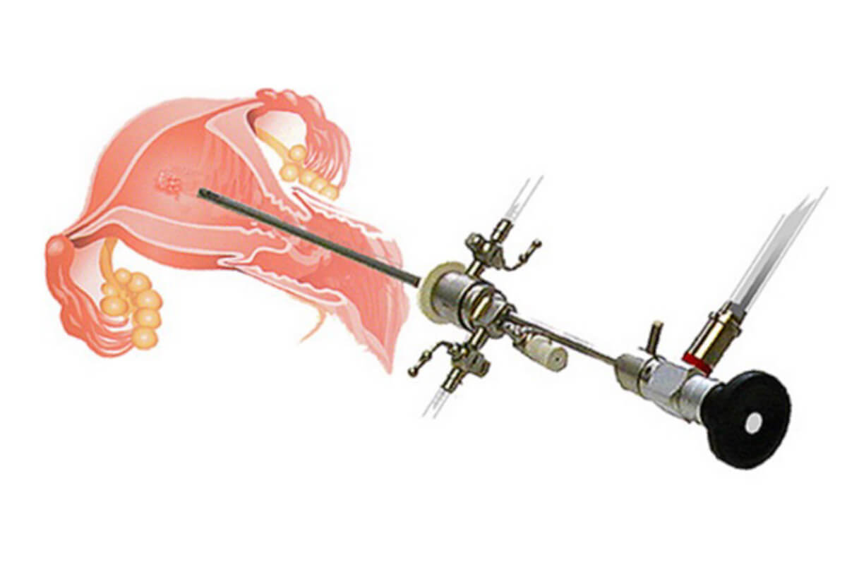
The procedure is performed with a camera attached to an optical fiber (light source). Other instruments, such as biopsy forceps, scissors, or resection loops, are also attached to the camera. By observing the images of the uterine cavity on a monitor, the doctor can diagnose lesions or remove them with surgical instruments.
Below are some images of our surgeries, highlighting the alterations commonly found in hysteroscopy.
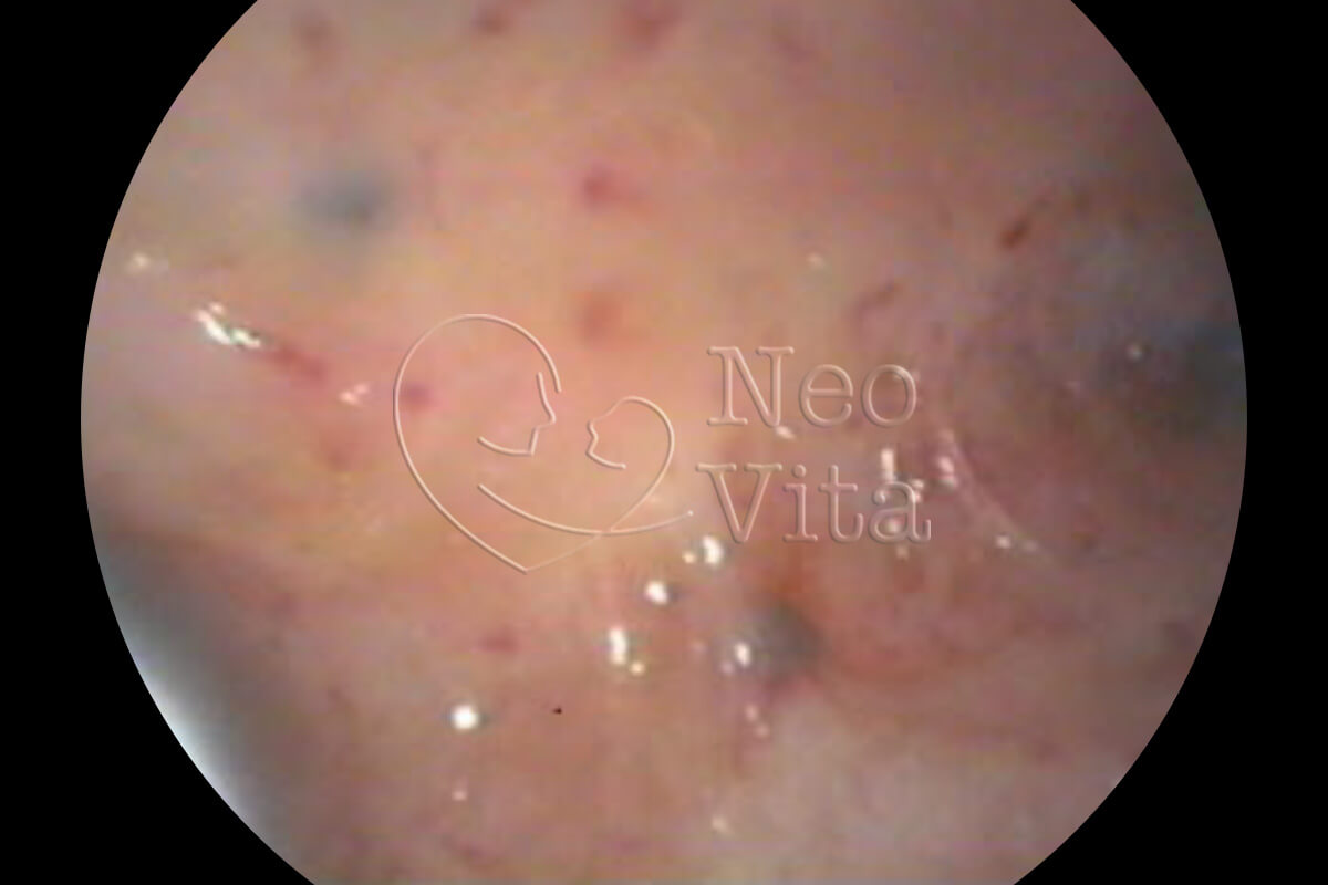
Note the darkened spots at the bottom of the uterus. These are typical invasions of adenomyosis.
The polyp is a projection of the membrane lining the uterus (the endometrium). It can cause bleeding or interfere with embryo implantation. They should be removed if identified in any previous imaging exam or during the hysteroscopy procedure.

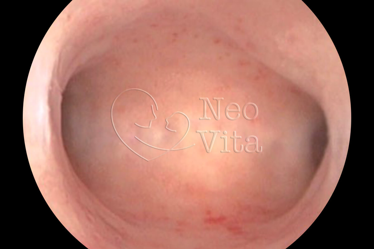
This is the portion of the uterus that connects the cavity to the uterine tube. Note that it is a narrow opening through which sperm must pass to meet the egg and fertilize it. It is not an alteration but an image showing the normal structure.
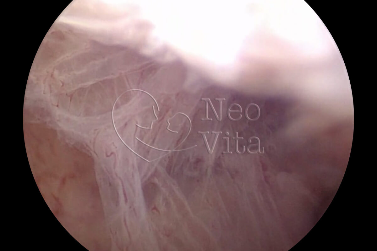
Synechia is the term used when a wall of the uterus adheres to another. They typically occur after uterine surgery, such as curettage, or even after childbirth. They must be undone so that the uterine cavity can return to its normal dimensions and allow pregnancy to develop.

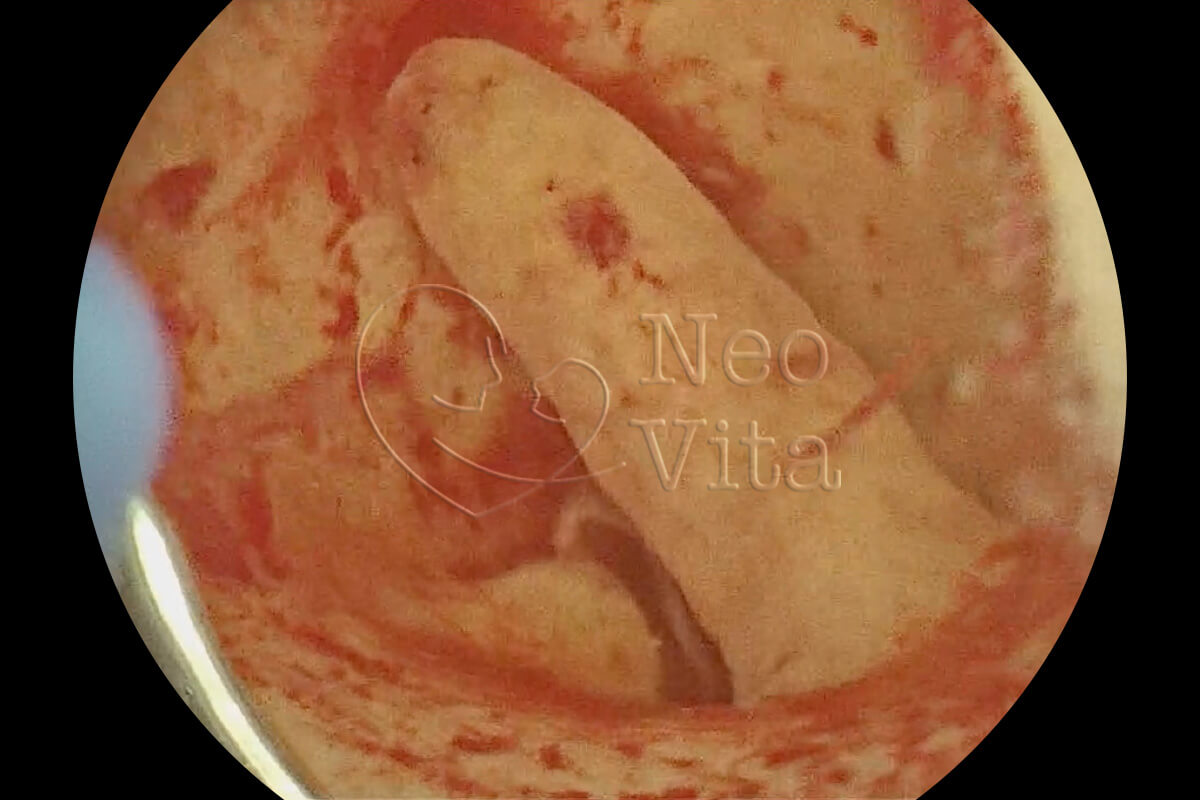
The photos show a fairly rare condition, acute endometritis. When we have chronic endometritis, the lesions are not as evident and are difficult to diagnose by hysteroscopy, recommending endometrial biopsy. In the case of acute endometritis, antibiotics should be administered.

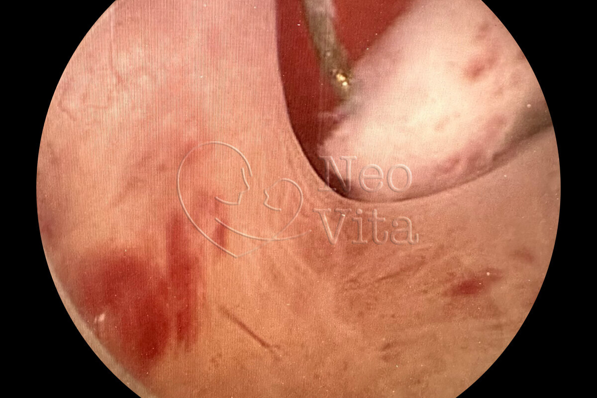
In the image, we see a submucosal fibroid and next to it, the resection loop used for the surgical removal of the lesion. Submucosal fibroids can cause bleeding, miscarriages, and even infertility.

The uterine septum is a fairly common malformation. Depending on its extent and thickness, it can lead to increased risks of early miscarriages. If there is a history of infertility, we recommend resecting the septum before embryo transfer.
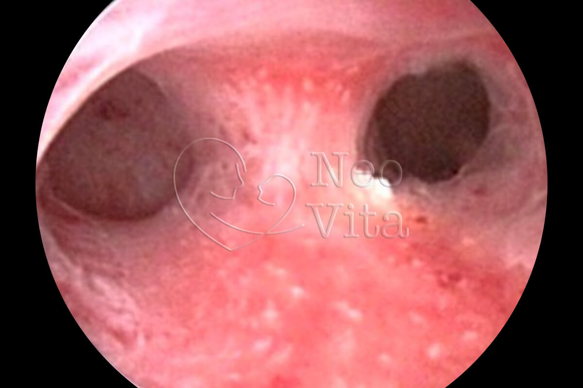
Here is another image showing a normal uterine cavity, with the uterine fundus in the center and the two tubal ostia on the sides.

Estamos aqui para oferecer um espaço acolhedor e informativo, onde você pode encontrar suporte, orientação e informações confiáveis ao longo da sua jornada.
Somos um
Que enfrenta os desafios da infertilidade e precisa da ajuda médica especializada para alcançar a tão sonhada gestação.
Ver tratamentos feitos para vocêSomos um
Que busca realizar o sonho da gestação através de uma jornada inclusiva e acolhedora.
Ver tratamentos feitos para vocêSou
Que deseja engravidar ou preservar a fertilidade para ser mãe no futuro.
Ver tratamentos feitos para vocêSou
Que deseja preservar a fertilidade ou engravidar para ser pai, com ajuda do tratamento Fertilização in vitro com ovodoação e barriga solidária.
Ver tratamentos feitos para vocêTratamentos personalizados para todos que desejam tornar seu sonho de ter filhos uma realidade.
 Português
  English